
The stethoscope, a vital tool in healthcare, has been a symbol of medical practice for over 200 years. Its design and functionality make it indispensable for doctors, nurses, and other healthcare professionals. Understanding the anatomy of a stethoscope and its components helps professionals use it effectively for accurate diagnoses. This guide explores the parts of a stethoscope, their specific functions, and best practices for optimal use.
Main Components of a Stethoscope
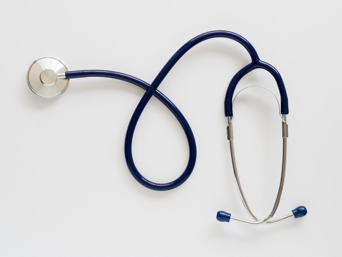
1. Chest Piece
The chest piece is the part of the stethoscope that comes into direct contact with the patient’s body. It consists of two sides:
- Diaphragm: The diaphragm is the larger, flat surface of the chest piece. It is designed to capture high-frequency sounds. For example, heartbeats, lung sounds, and bowel activity. Its structure allows for a broad range of auscultations, making it essential for general diagnostics.
- Bell: The bell is the smaller, concave side of the chest piece. It is specifically used to detect low-frequency sounds. For instance, vascular murmurs and bruits. This feature is particularly helpful for pediatric patients or when diagnosing faint sounds that the diaphragm might miss.
When to Use Bell vs. Diaphragm:
- Use the diaphragm for most routine examinations, such as checking heart and lung sounds in adult patients.
- Opt for the bell when you need to detect subtle, low-frequency sounds or during pediatric assessments.
| Feature | Diaphragm | Bell |
| Use | High-frequency sounds | Low-frequency sounds |
| Example | Heartbeats, lung sounds | Bruits, heart murmurs |
| Technique | Press firmly on the skin | Press lightly on the skin |
Open vs. Closed Bell:
- An open bell works best for amplifying low-pitched sounds. It is often used for detecting abnormal vascular sounds.
- A closed bell may reduce background noise but can slightly limit sound sensitivity.
| Aspect | Open Bell | Closed Bell |
| Design | Separate bell | Integrated with diaphragm |
| Use | Low-frequency sounds only | Switch between high and low sounds |
| Convenience | Requires manual switching | More versatile |
2. Tubing
The tubing connects the chest piece to the headset. It is a flexible, hollow tube that transmits sound waves from the chest piece to the earpieces. High-quality tubing minimizes sound loss and external interference, ensuring accurate sound transmission.
Key Features of Stethoscope Tubing:
- Durability: Thick, latex-free tubing is preferred for longevity.
- Length: Standard tubing length ranges from 22 to 28 inches, allowing for comfortable use during examinations.
3. Headset
The headset comprises the metal ear tubes and the earpieces. It is designed to fit comfortably in the user’s ears while maintaining optimal sound transmission. The headset’s spring tension can often be adjusted to ensure a snug but comfortable fit.
4. Earpieces
Earpieces are soft, rubber, or silicone tips that fit snugly into the ears. They create an airtight seal, which is critical for isolating ambient noise and accurately transmitting body sounds.
Tips for Earpiece Maintenance:
- Clean regularly to prevent ear infections.
- Replace worn or hardened earpieces to maintain sound quality and comfort.
5. Stem
The stem is a small metal component that connects the chest piece to the tubing. It often contains a rotating mechanism, allowing users to switch between the diaphragm and the bell.
6. Acoustic Valve
This valve regulates sound flow and toggles between the diaphragm and bell functions. It ensures sound clarity and prevents loss of acoustic details during examinations.
Functions of a Stethoscope
A stethoscope serves as an essential diagnostic tool for medical professionals. Its applications include:
- Measuring Heart Rate and Rhythm: Detect arrhythmias or irregular heartbeats.
- Lung Examinations: Identify abnormal lung sounds such as wheezing, crackles, or diminished breath sounds.
- Blood Pressure Measurement: When paired with a sphygmomanometer, it detects Korotkoff sounds.
- Bowel Sound Analysis: Helps detect gastrointestinal obstructions or hyperactive sounds.
- Vascular Sound Detection: Identifies bruits and other vascular abnormalities.
- Fetal Heart Monitoring: Common in prenatal care to detect fetal heartbeats.
- Joint Examination: Monitors joint sounds, especially in conditions like temporomandibular joint dysfunction.
- Thoracic Trauma Assessment: Checks for irregularities in lung function or chest injuries.
- Heart Murmur Diagnosis: Detects abnormal heart sounds caused by valve issues.
- Post-Surgical Recovery: Monitors organ function during recovery periods.
Bell Vs Diaphragm: Key Differences
Understanding the differences between the bell and diaphragm enhances diagnostic accuracy.
| Feature | Bell | Diaphragm |
| Frequency | Low-frequency sounds | High-frequency sounds |
| Best For | Murmurs, vascular sounds | Heart and lung sounds |
| Use Case | Specialized, pediatric applications | General adult diagnostics |
Comparing Stethoscope Vs Sphygmomanometer Parts
Stethoscopes and sphygmomanometers are two essential tools in medical diagnostics, often used together during patient examinations.
Each has distinct parts and functions that contribute to accurate assessments of a patient’s health. Below is a detailed comparison of their components and purposes.
| Component | Stethoscope | Sphygmomanometer |
| Chest Piece/Diaphragm | Detects Korotkoff sounds and body noises | Not applicable |
| Tubing | Transmits sound from chest piece to earpieces | Connects cuff to the pressure gauge |
| Earpieces | Used for listening to body sounds | Not applicable |
| Cuff | Not applicable | Wraps around the arm to restrict blood flow |
| Gauge | Not applicable | Measures systolic and diastolic pressures |
| Valve | Not applicable | Regulates air release from the cuff |
Key Roles
- Stethoscope: Primarily used for auscultation, it amplifies sounds such as heartbeats, lung functions, and Korotkoff sounds during blood pressure measurement.
- Sphygmomanometer: Measures blood pressure by inflating a cuff around the arm and detecting changes in arterial blood flow.
Integration in Diagnostics
When used together, the stethoscope and sphygmomanometer provide comprehensive insights into cardiovascular health. For instance, the stethoscope detects Korotkoff sounds as the sphygmomanometer records pressure readings, enabling precise systolic and diastolic measurements.
Understanding the specific roles and components of these tools ensures accurate diagnosis and effective patient care.

Maintenance Tips for Stethoscopes
Proper care ensures the longevity and effectiveness of your stethoscope:
- Clean the Chest Piece: Wipe with alcohol after each use to prevent cross-contamination.
- Avoid Extreme Temperatures: Heat and cold can degrade the tubing.
- Store Properly: Keep the stethoscope in a clean, dry place away from sharp objects.
- Check Earpieces Regularly: Replace as needed to maintain comfort and hygiene.
- Inspect Tubing for Cracks: Replace damaged tubing to ensure sound quality.
Read More: Who Invented the Stethoscope? A Look at René Laennec’s Legacy
Conclusion
The stethoscope is more than a diagnostic tool; it is a gateway to understanding a patient’s health. By mastering its anatomy and functions, healthcare professionals can improve diagnostic precision and patient care.
From the differences between the bell and diaphragm to the importance of maintenance, understanding the stethoscope’s components is essential for every practitioner.
For additional resources, explore the links and references provided to deepen your knowledge of this indispensable medical instrument.
