
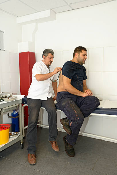
Auscultation is a key part of a physical examination. It allows you to listen to internal body sounds using a stethoscope, especially the heart and lungs. Whether you’re a healthcare professional or a student, learning common stethoscope auscultation techniques is essential for detecting abnormalities and diagnosing conditions early.
What is Auscultation?
Auscultation means listening to the sounds made by the heart, lungs, and other organs to assess their health. This involves identifying normal and abnormal sounds, which can reveal conditions like heart murmurs, lung infections, or fluid buildup.
Lung Auscultation Techniques
Auscultation Sites of the Lungs
The lungs have specific areas for auscultation. These are divided into anterior (front), posterior (back), and lateral (sides). Follow these landmarks to ensure a thorough exam:
Anterior Chest
- Start at the trachea for bronchial sounds.
- Move to the upper lobes of both lungs (above the clavicles).
- Continue to the middle zones and lower areas near the ribs.
Posterior Chest
- Begin at the top near the shoulders.
- Listen to the interscapular regions (between shoulder blades).
- Move downward to the lower back regions, where the base of the lungs sits.
Lateral Chest
- Listen to the sides of the chest.
- Focus on the middle and lower lung zones.
Tip: Ensure the patient breathes deeply through their mouth for better sound clarity.
Common Lung Sounds
During auscultation, you will encounter normal and abnormal lung sounds:
| Type | Description | Significance |
| Vesicular | Soft, low-pitched sounds during inhalation and exhalation. | Normal lung sounds. |
| Crackles (Rales) | Discontinuous popping or crackling noises. | Fluid in the airways (e.g., pneumonia). |
| Wheezes | High-pitched, musical sounds during exhalation. | Narrowed airways (e.g., asthma or COPD). |
| Rhonchi | Low-pitched, snoring-like sounds. | Mucus or obstruction in larger airways. |
| Stridor | Harsh, high-pitched sound during inhalation. | Upper airway obstruction. |
Key Tips for Lung Auscultation
- Use the diaphragm of the stethoscope for high-pitched lung sounds.
- Compare both sides of the chest to detect asymmetries.
- Note any absence of sounds, which might indicate a collapsed lung.
Heart Auscultation Techniques
5 Areas of the Heart for Auscultation
The heart has five key areas where sounds are best heard. These areas correspond to specific valves and regions of the heart.
Aortic Area:
- Located at the right second intercostal space, near the sternum.
- Used to hear sounds from the aortic valve.
Pulmonic Area:
- Found at the left second intercostal space.
- Ideal for listening to the pulmonic valve.
Erb’s Point:
- Located at the left third intercostal space.
- A good spot to hear S1 and S2 heart sounds equally.
Tricuspid Area:
- Found at the left fourth or fifth intercostal space near the sternal border.
- Best for hearing the tricuspid valve.
Mitral Area (Apex):
- Located at the left fifth intercostal space near the midclavicular line.
- Perfect for detecting mitral valve sounds.
| Area | Primary Valve | Common Sounds Heard |
| Aortic | Aortic Valve | Murmurs, S2 sounds. |
| Pulmonic | Pulmonic Valve | Ejection clicks, murmurs. |
| Erb’s Point | Both Aortic & Pulmonic | Equal S1 and S2 sounds. |
| Tricuspid | Tricuspid Valve | S1 sounds, low-pitched murmurs. |
| Mitral | Mitral Valve | S1 sounds, gallops, and murmurs. |
Auscultation of Heart Sounds
Heart sounds are categorized into normal and abnormal types:
- S1 (Lub): Caused by the closure of mitral and tricuspid valves.
- S2 (Dub): Caused by the closure of aortic and pulmonic valves.
- Abnormal sounds:
- Murmurs: Result from turbulent blood flow.
- Gallops: Extra sounds (S3 or S4) linked to heart failure.
- Clicks: Indicate valve issues (e.g., mitral valve prolapse).
Pro Tip: Use the bell of the stethoscope for low-pitched heart sounds like S3 or S4.
How to Perform Auscultation in Physical Examination
When performing auscultation in physical examination, it’s crucial to follow these steps:

Prepare the Patient:
- Ensure the patient is in a quiet environment.
- Position them sitting up or lying down as needed.
Use the Stethoscope Properly:
- Use the diaphragm for high-pitched sounds (e.g., lung or S1/S2 heart sounds).
- Use the bell for low-pitched sounds (e.g., S3/S4 or murmurs).
Compare Bilaterally:
- Always check both sides of the chest or back to identify differences.
Document Findings:
- Record your observations immediately to avoid missing details.
Comparison of Heart and Lung Auscultation
| Aspect | Lung Auscultation | Heart Auscultation |
| Primary Tool | Diaphragm of stethoscope | Diaphragm and bell of stethoscope |
| Focus Areas | Front, back, and sides of the chest | 5 key areas (aortic, pulmonic, etc.) |
| Common Sounds | Vesicular, crackles, wheezes | S1, S2, murmurs, gallops |
| Purpose | Detect lung issues like pneumonia or asthma | Identify heart abnormalities like murmurs or valve issues |
Conclusion
Mastering common stethoscope auscultation techniques is a must for accurate physical examinations. Whether you’re checking the auscultation sites of the lungs or the 5 areas of the heart, practice, and attention to detail are key. Recognizing normal and abnormal auscultation sounds ensures you catch issues early, improving patient outcomes.
